Silicified coral with transparent chalcedony filling corallite - Indonesia
- Details
- Created: Monday, 22 July 2024 21:44
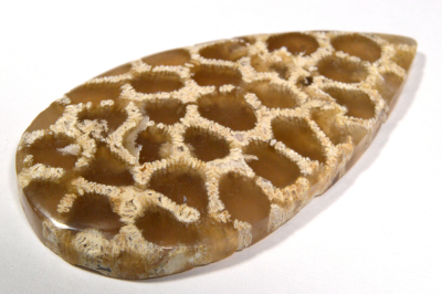
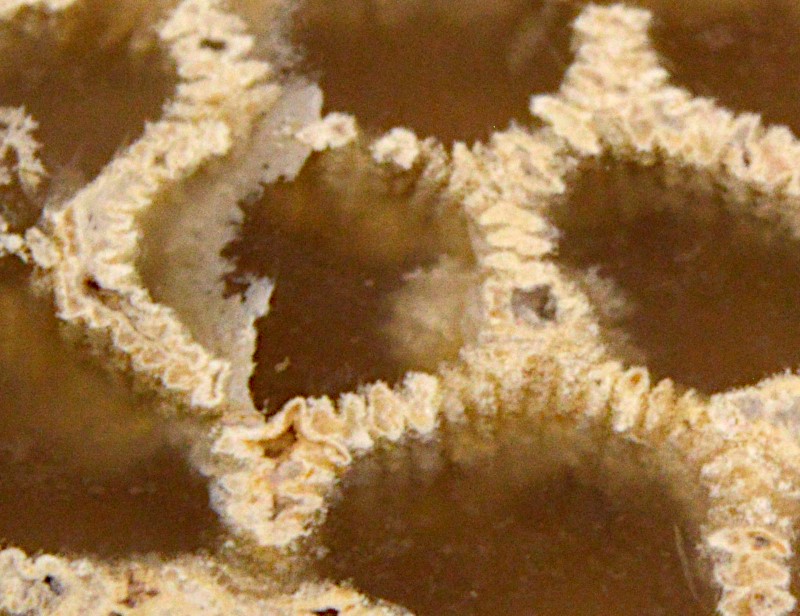
The fossilized corals have been quite common in the gemstone market for years. Most of the time, the material is opaque and almost all the structure / anatomy of the calcium carbonate skeletons of the corals is preserved. Each individual coral 'cell' hosting the polyp (the living part), known as the corallite, forms a kind of cylinder (calyx) delimited by a wall (theca) that contains a columnar framework based on the septa. Seen from above, the septa are like the radius of a bicycle wheel. In the presented cabochon (Figure 1), this 'internal' framework is missing, only the wall and septacostae (small septa across the wall) remain. Most likely the silification (filling with and / or transforming to siliceous minerals) occurred long after the coral dyed and its internal structure got destroyed or dissolved. Visually, the cabochon looks like a honeycomb structure. Details of the corallite can be better seen in Figure 2.
 Figure 1. The 26.75 ct cabochon of a coral fossil silicified with
transparent chalcedony filling the calyx of the corallite (coral's
cylinder-like structure).
| Shape | pear cabochon |
| Size | 40.4 x 21.6 x 4.6 mm |
| Color | honey filling corallite, gray to white septacostae and walls |
| Diaphaneity | transparent within corallite, opaque for septacostae and walls |
| Lustre | vitreous |
| Weight | 27.65 ct |
| SG | ~ 2.0 (the cabochon is partly hydrophane, it absorbs approximately 0.5 ct of water in few seconds) |
| RI | 1.540 (transparent area from the back) |
| DR | - |
| Pleochroism | - |
| Polariscope / Conoscope | transparent area stay light through a full 360° rotation -> polycrystalline |
| SWUV | inert |
| LWUV | inert |
| Magnetic susceptibility N52 | very slightly attracted although chalcedony is usually diamagnetic, likely due to some other minerals / impurities |
| Chelsea filter | inert |
Table 1. Observational and measured properties

Figure 2. Closer view to the corallite and its calyx filling with honey brown chalcedony as demonstrated by spectroscopy. The view's width
corresponds to about 10 mm.
Infrared reflectance spectroscopy:
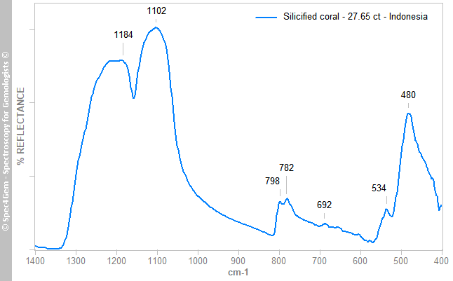
The Infrared reflectance spectrum (Figure 3) was collected from a honey brown area located on the top of the cabochon. The reflectance bands at 480, 534, 692, 782, 798, 1102 and 1184 cm-1 characterize α-quartz especially cryptocrystalline chalcedony (the 782 and 798 cm-1 bands are still well defined if compared to opal but far less sharper than these of a quartz monocrystal).
 Figure 3. The IR reflectance spectrum of the honey brown material shows reflectance bands at 480, 534, 692, 782, 798, 1102 and 1184 cm-1 that characterize α-quartz especially cryptocrystalline chalcedony.
Raman spectroscopy:
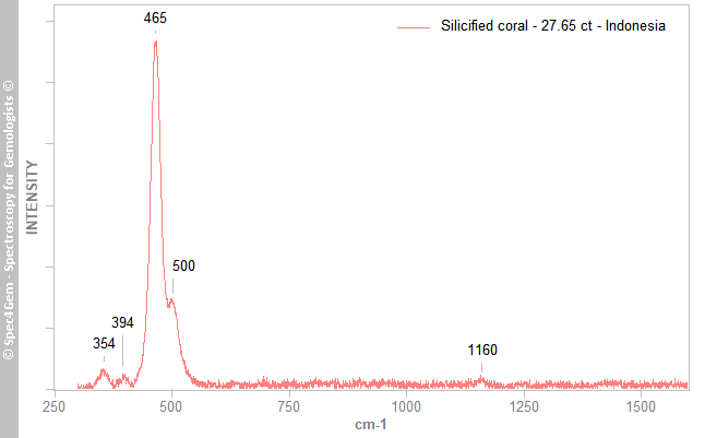
The Raman spectrum (Figure 4) was collected from a honey brown area located on the top of the cabochon exactly as for IR spectrum. The 464 cm-1 prominent peak with the additional peaks at 354, 394 and 1160 cm-1 characterize quartz and the presence of the 500 cm-1 related to moganite indicates that the honey brown material is chalcedony.
 Figure 4. The Raman spectrum of the honey brown material shows peaks at 354, 394, 465, 500 and
1160 cm-1. With the prominent peak at 465 cm-1 which is related to quartz and the 500 cm-1 peak related to moganite, the honey brown colored material is chalcedony.
UV-VIS-NIR spectroscopy:
No investigation of the honey brown color of the chalcedony.
Photoluminescence spectroscopy:
As the cabochon is inert to both 254 and 365 nm excitations, no further investigation was performed.
Under the 365 nm excitation and microscope, there are many spots located at the interface between the walls and the honey brown chalcedony filling the calyx that glow bright red. It is possibly another mineral or a contaminant.
Conclusion:
This cabochon represents the case of a coral fossil that was silicified with the filling of its corallites with transparent honey brown chalcedony, most of its septa disappeared except the septacostae that are placed across the theca (wall). With its transparency this cabochon differs from most of other coral fossil cabochon from Indonesia.
Further investigations could be conducted to identify the nature of the septacostae / wall.

