
Multicolored fluorite prism with white inclusions supposedly from China
- Details
- Created: Tuesday, 07 June 2022 21:02
Many fluorite prisms have been available for years. These are natural fluorite material shaped and polished like a hexagonal prism terminated by a pyramid and by either a second pyramide or a basal plane. The fluorite photographed in the Figure 1 is green and violet with some white inclusions. Sometime the prism is made of banded fluorite and it is accordingly named rainbow fluorite.
The white feathery inclusions are very common in those fluorites and they have been subject to many interrogations. Many information circulating over internet suggested they could be scolecite (a zeolite). A discussion in the Identity Help section hosted by Mindat[1] stated from qualitative EDX analysis that Zn, Si, O elements were detected and the white material could be a zinc silicate. Another proposition was that it could be calcite in the paper spar variety (with a highly compressed habit) or a quartz pseudomorphose after it.
In the sample from the present study, the inclusions are mainly located in two zones, one in the green fluorite pyramid terminating the prism (Figure 2), and the other one in the middle of the prism in the violet fluorite that seemed to grow on a plane corresponding to a fluorite face (Figure 3). Although the feathery inclusions in the green part are well formed, the ones in the violet part look to be partially dissolved.
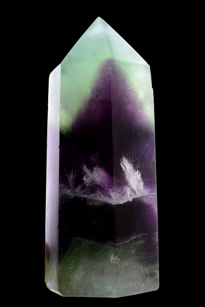 Figure 1. The 84 grams hexagonal prism of bi-colored fluorite
 showing feathery white inclusions.
| Shape | Hexagonal prism terminated by a pyramid and a basal plane, man made shape |
| Size | 80 x 43 x 26 mm |
| Color | Green, violet and white inclusions |
| Luster | Dull to vitreous |
| Weight | 84 g |
| SG | - |
| RI | - |
| DR | - |
| Pleochroism | None, fluorite is isotropic |
| Polariscope / Conoscope | Isotropic |
| SWUV | Bluish for the green fluorite, strong yellowish-white to white for the white inclusions |
| LWUV | Weak bluish for the green fluorite, strong yellowish-white to white for the white inclusions |
| Magnetic susceptibility N52 | Diamagnetic (repulsion : 81 g under the magnet although it is 84 g without) |
| Chelsea filter | Inert for fluorite but the inclusions appear white contrasting with the fluorite |
Table 1. Observational and measured properties
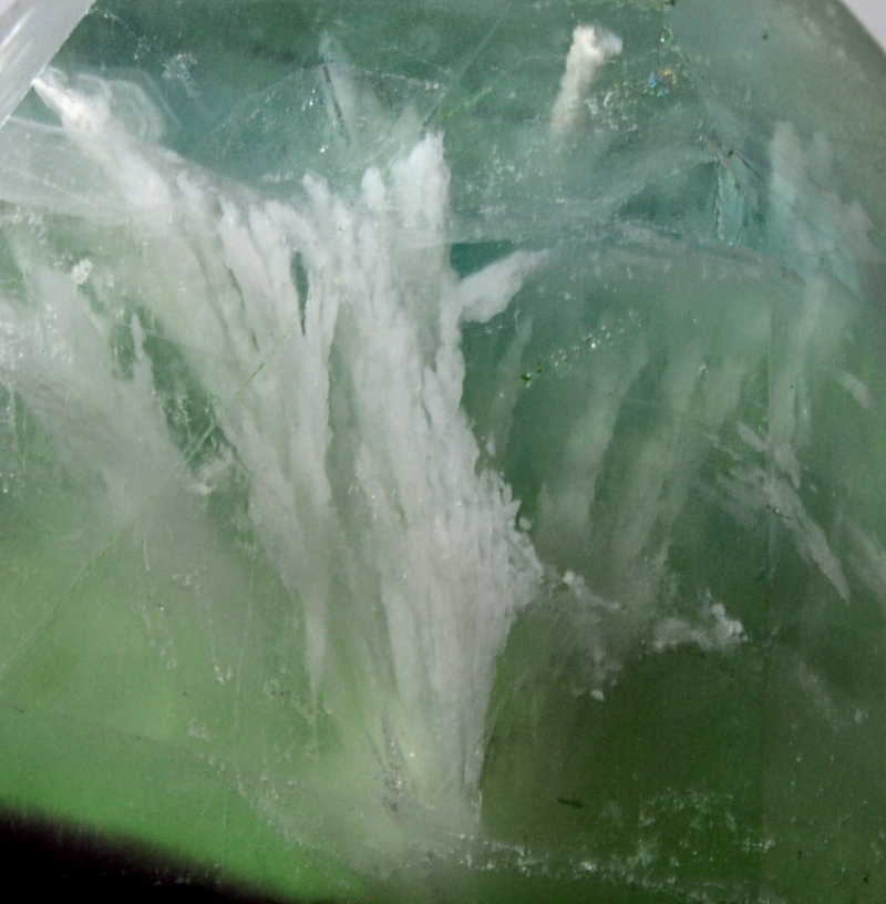
Figure 2. The pyramid end contains 'feathery' or 'thuja coniferous leaves' white inclusions, all starting from a common point. They are
up to one centimeter long and some are flush to pyramid faces. Photographed with a 55 mm digital camera lens.
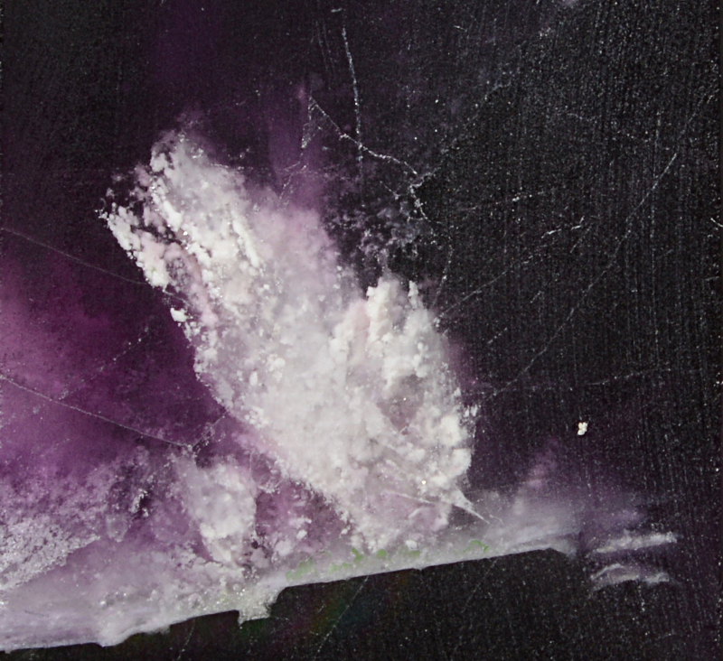
Figure 3. The inclusion scene in the violet fluorite significantly differs in appearance from that observed in the green fluorite pyramid. The
inclusions look like more disjoint snow flakes but a close observation shows they have finally the same habit than the feathery ones in
the green pyramid except they have been partially dissolved. Photographed with a 55 mm digital camera lens.
Infrared reflectance spectroscopy:
Infrared reflectance spectra were collected from three points (small areas), in the green and violet part without any white inclusion (Figure 4), in the pyramid part where the inclusions flush with the surface (Figure 5) and in the violet part where the inclusions flush with the surface but in a lesser extend than in the pyramid (Figure 7).
The Figure 4 spectrum indicates fluorite with a reflectance singularity on the 400 - 500 cm-1 range where the reflectance is lower than at 500 cm-1. Typical fluorite spectra reflectance is as shown in Figure 6 for reference.
The Figure 5 spectrum cannot not be matched to a single mineral, two bands at 781 and 798 cm-1 are easily recognized as being part of a quartz spectrum (see Figure 6 for a quartz reference spectrum). The other narrow bands at 611, 648 and 978 cm-1 are found to match those of baryte, a barium sulfate formed in hydrothermal depositional environments, the baryte reflectance spectrum is shown in Figure 6. The broad band from 400 to 600 cm-1 is similar to that of the characteristic band of fluorite. To illustrate the collected composite spectrum, three spectra of fluorite, baryte and quartz as shown in Figure 6 were summed, the resulting spectrum is shown in Figure 5 for comparison with the collected spectra, the match is convincing. The strong band of quartz (400 - 500 cm-1) added to that of fluorite creates a reflectance spectrum similar to that observed for the fluorite body, possibly contaminated with quartz.
The Figure 7 spectrum cannot be confused with any other mineral than quartz, it shows a crystalline quartz reflectance spectrum. At the spectrum collection point in the white inclusion area of the violet fluorite, quartz is significantly present at the surface. Unlike in the green pyramid, the baryte cannot not be detected, can it be alongside ? The FTIR reflectance setup does not allow to easily hit a particular point, especially with such big specimen.
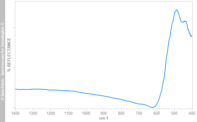 Figure 4. IR reflectance spectrum of the prism body, green and violet areas. The spectrum indicates fluorite with an unusual reflectance on the 400 - 500 cm-1 range for the fluorite, the reflectance on this range is usually higher than that at 500 cm-1.
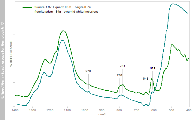 Figure 5. IR reflectance spectrum of the white inclusions flushing with the surface of the green pyramid (Figure 2). The spectrum does match directly any known spectrum. The green spectrum named "fluorite 1.37 + quartz 0.93 + baryte 0.74" is issued of the sum of the three spectra given for reference in Figure 6, it shows the composite spectra of fluorite, quartz and baryte all together and it amazingly matches the sample's spectrum. The 611, 648 and 978 cm-1 bands are indicative of baryte and the 781 and 798 cm-1 ones are those of quartz.
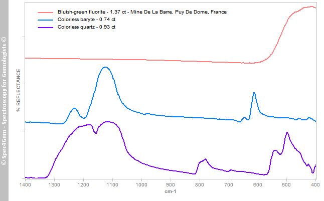 Figure 6. IR reflectance spectra (from top to bottom, shifted for clarity) of a fluorite, a baryte and a quartz. These three spectra were summed to produce the composite spectrum given in Figure 5.
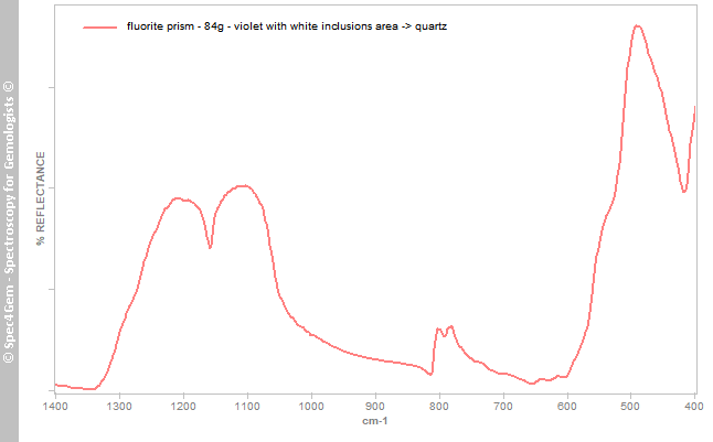 Figure 7. IR reflectance spectrum of the the white inclusions area in the violet part (Figure 3). At this collection point, the spectrum is undoubtedly that of quartz.
Raman spectroscopy:
The power and the focusing system of a Raman equipment makes possible to reach inclusions under the surface. The Raman spectrum of the white inclusions of the violet fluorite part (Figure 3) using a 636 nm laser is shown in the Figure 8 and it indicates the white inclusions mineral is baryte and it confirms what is observed in the green pyramid with the FTIR. Raman spectra can be found at RRUFF site[2] for reference.
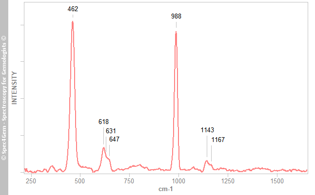 Figure 8. Raman spectrum (with a 636 nm laser) of the white inclusions in the violet fluorite as photographed in Figure 3. The Raman bands at 462, 618, 631, 647, 988, 1143 and 1167 cm-1 indicate baryte as the mineral.
UV-VIS-NIR spectroscopy:
No UV-VIS-NIR spectroscopy study has been conducted on this sample since the main goal was to identify the white inclusions.
Photoluminescence spectroscopy:
No photoluminescence spectroscopy study has been conducted on this sample since the main goal was to identify the white inclusions.
Conclusion:
As the FTIR reflectance and Raman spectroscopies studies show, the white mineral included as "feather / coniferous leaves / snow flakes" is baryte, the barium sulfate. The dissolved white inclusions in the violet fluorite part are also accompanied by quartz.