
Ophite from Urals mountains, Russia
- Details
- Created: Monday, 31 December 2018 10:58
With a quite strong light it is possible to observe by transparency some darker green veins with surrounding reddish areas. This phenomenom has already been observed in Cr- antigorite and bowenite showing the usambara effect, here the phenomenom is rather weak. The gemstone is viewed red through the chelsea filter if the stone is backlighted with an incandescent light.
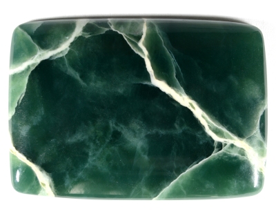 Figure 1. The 70 ct cabochon of dark-grayish-green ophite from
Urals mountains, Russia, shows some whitish areas and few white
thin veins.
| Shape | flat rounded corners rectangular cabochon |
| Size | 45.6 x 32.1 x 4.05 mm |
| Color | dark-grayish-green shades with some white veins |
| Lustre | greasy |
| Weight | ~ 70 ct |
| SG | |
| RI | - |
| DR | - |
| Pleochroism | - |
| Polariscope / Conoscope | stays light |
| SWUV | the white veins are white and the green material is inert |
| LWUV | the white veins are strong white and the green material is inert |
| Magnetic susceptibility N52 | quite strong, the N52 magnet drags the stone |
| Chelsea filter | inert in reflected light while it is red in transmitted incandescent light from the back |
Table 1. Observational and measured properties
Infrared reflectance spectroscopy:
The IR reflectance spectrum (figure 2) was collected from the cab's top in a green area. The reflectance equipment used to collect does not allow to get the reflected IR from the white veins since they are embbeded in the green material and they cannot be isolated. The spectrum's pattern is characteric of serpentines. It is not worth to speculate in identifying the three serpentines species (antigorite, chrysotile and lizardite) within the spectrum. The 568 cm-1 band generally attributed to Mg-O is characteristic of antigorite.
 Figure 2. IR reflectance spectrum of a green area of the ophite cab showing a characteristic serpentine pattern.
UV-VIS-NIR spectroscopy:
The UV-Vis-NIR spectrum (figure 3) was collected from a green area of the cab with the light path travelling from the top to the back of the cab. It shows a hypothetical strong absorption around 400 nm followed by another quite stong band at 572 nm. The asymetry of the 572 nm band suggest another band around 630 nm. A small feature at 685 nm is possibly connected to Cr3+, but it is not clear here if it is an absorption band or a luminescence artefact. There is a weak and large band centered at 730 nm followed by another large band peaking around 905 nm. As in most of the serpentines, the triplet at 946, 954 and 961 nm is well visible in the spectrum.
Ni2+, Fe2+ serpentines show large absorptions in the NIR range, except the 730 nm band, the almost unexistant 905 nm band, the gemstone is likely not colored by iron and nor nickel even if they can be present. The 572 nm and the secondary 685 nm small feature likely indicates a possible coloration by Cr3+. The too strong absorption in the violet cannot be collected because of spectrophotometer's limits, but as written earlier, the presence of the hypothetical strong absorption around 400 nm could confirm the Cr3+ as the color cause.
 Figure 3. The UV-Vis-NIR spectrum collected from a dark-green area shows two humps, one hypothetical around 400 nm and the second one at 572 nm likely attributed to Cr3+ as well as the 685 nm one. The weak features at 630, 730 and 905 nm are possibly related to Fe2+ and the triplet at 946, 954 and 961 is still to be discovered.
Photoluminescence spectroscopy:
The luminescence spectroscopy was performed with the 405 nm laser, two areas were investigated, a green area and a white vein, the spectra are shown in figure 4. The green area shows a red luminescence with a large emission centered around 730 nm which is decorated with three small peaks at 692, 708 and 727 nm. Such red emission is quite characteristic of Cr3+. The white vein shows a white luminescence as observed with SWUV and LWUV lamps, which is characterized by a large emission between 400 and 650 nm with a maximum around 510 nm. Could it be organic molecules luminescence, further studies have to be conducted to understand it, knowing the nature of the white material might easily explained it.
 Figure 4. The photoluminescence spectrum of the green area shows Cr3+ red emission (692, 708, 727 nm). The white veins show a large white emission between 400 and 650 nm peaking at 510 nm, this emission is unexplained at the moment.
Conclusion:
The green material is serpentine as confirmed by IR spectroscopy but the mixture of the three serpentine varieties entering in the composition (antigorite, chrysotile and lizardite) has not been investigated. The white veins material was not investigated as well and it is still unknown. The color is likely caused by Cr3+ as shown by UV-Vis spectroscopy and those presense is confirmed by the red photoluminescence. Fe2+/Ni2+ are possibly present but likely in weak concentration and do not affect the NIR spectrum. The Cr3+ and Fe2+/Ni2+ can also explain the gemstone strong reaction to the N52 magnet.
This ophite material from Russia is visually interesting although is not often seen in the market.