
Long mistaken for green peridot, dunilite was identified in 2000 as a new gem variety from olivine group but not yet approved by IMA. It is an orthorhombic, magnesium, iron, manganese silicate. Though the x-ray diffraction pattern shows that the line spacing is similar to peridot. These spacing, however are not identical with those for peridot (By Dr Mary L Johnson, GIA July 8 1998).
The colour of dunilite varies from light greenish yellow to brownish green, the depth of colour being related to the iron content. Dunilite is not quite as hard as quartz. It does not however, have cleavage.
Dunilite occurs in only one locality called, “Katukubura mountain” Kolonne, Sri Lanka. It has been found as tabular rectangle shaped crystals in white calcite rock. The first morphological data for dunilite was obtained from a 0.97-carat faceted gemstone from GIA 10318180 Dated March 13th, 1998 confirms the stone as a new member of the olivine group.
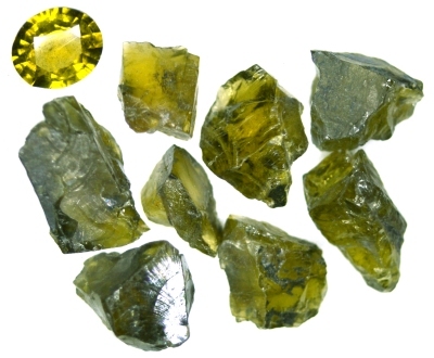 Figure 1. 0.26 ct faceted stone and and 5.73 ct of rough dunilite.
| Shape | 1 brilliant / step cut oval stone and 8 rough stones |
| Size | 4.44 x 3.84 x 2.13 mm |
| Color | brownish green-yellow |
| Lustre | vitreous |
| Weight | 0.26 ct for the cut stone |
| SG | 3.49 |
| RI | 1.679 - 1.716 |
| DR |
0.037 B - |
| Pleochroism | grayish-green, brownish-yellow |
| Polariscope / Conoscope | anisotropic, biaxial |
| SWUV | inert |
| LWUV | inert |
| Magnetic susceptibility | strong (N52 magnet picks up the 1.15 ct rough stone) |
Table 1. Observational and measured properties
Infrared reflectance spectroscopy:
The IR reflectance spectrum was acquired from the table of the 0.26 ct cut stone.
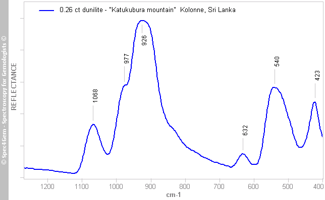
Figure 2. IR reflectance spectrum acquired from the table of the 0.26 ct cut stone.
UV-VIS-NIR spectroscopy:
The UV-Vis-NIR absorption spectrum was acquired with a light path going from the culet to the table, thus perpendicular to the table.
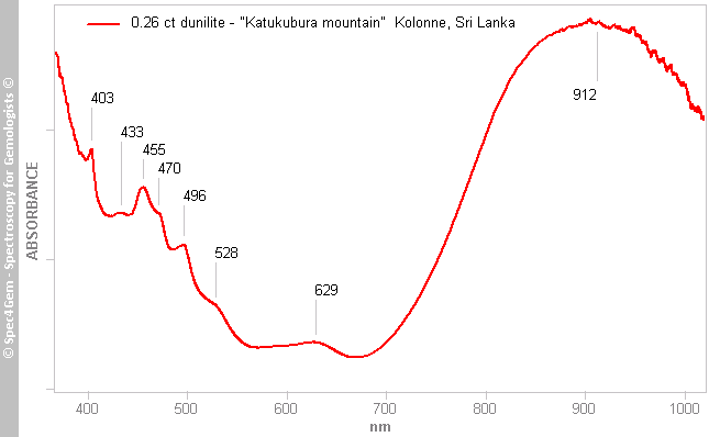
Figure 3. Caption of above spectrum
Photoluminescence spectroscopy:
No photoluminescence observed with 254 nm and 405 nm excitation. This is likely caused by the presence of Fe2+.