
A small dark violetish-blue sapphire with a strong color saturation weighing 0.84 ct has raised a lot of questions. Especially the question to know if this sapphire is a natural or synthetic one.
This sapphire, shown in figure 1, was submitted to a classical gemology analysis and to some spectroscopic analysis to try to answer to the question.
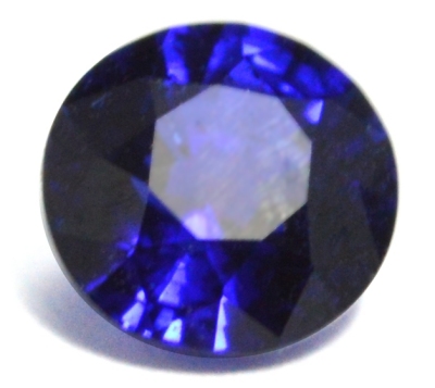 Figure 1. 0.84 ct dark violetish-blue sapphire.
| Shape | brilliant round |
| Size | ∅ 5.47mm x 3.45 mm |
| Color | dark violetish-blue, homogeneous, no color zoning |
| Lustre | bright vitreous |
| Weight | 0.84 ct |
| SG | ~ 3.9 [3.95 - 4.05] |
| RI | 1.760 - 1.769 [1.760 - 1.775] |
| DR | 0.009 Uniaxial - [0.008 - 0.009, U-] |
| Pleochroism | strong violetish-blue (down to table, || to C) / greenish-blue (_|_C) |
| Polariscope / Conoscope | table || filters -> uniaxial interference figure (cross) seen through the culet : table _|_C table _|_ filters -> light and dark four times per 360° rotation |
| SWUV | inert |
| LWUV | inert |
| Magnetic susceptibility | inert |
Table 1. Observational and measured properties
The stone presents numerous inclusions that can be described as tiny bubble included in a slightly larger bubble, a kind of two phase inclusion, the included bubble does not seem to move in is host bubble. The bubble is generally regular (sphere), sometimes a bit elongated. Some of these bubble inclusions are close or linked to long flat irregular kind of 'arrow' black inclusions. Such bubbles could be glass fluid inclusions that appears also in natural sapphires, especially in Montana sapphires. It is also important to note that there is no color zoning, color is homogeneous through all the stone.
Observational and measured properties summarized in table 1 can be interpreted as is:
- The RI are low and seem to indicate a low iron content (iron concentration is the main factor modifying the RI). The interference figure observed down to the table ( || C) within the polariscope is easily seen without the aid of the conoscope.
- The pleochroism, strong violetish-blue || C and greenish-blue _|_C, is strong but the main characteristic is that the pleochroism is inverted compared to that of the common natural blue sapphires (light blue || C and strong blue _|_C). Such "inverted" pleochroism is known for Russian hydrothermal synthetic sapphire mainly colored by Ni2+ [1].
- There is no observable luminescence while the stone is exposed to both LWUV and SWUV, either the UV sources are too weak to excite the luminescence or there is no chromophore nor center to respond to the excitation or the luminescence is quenched by some ions as Fe2+.
- The stone does not show any magnetic susceptibility since it is totally inert to the N52 magnet, likely because iron content is too weak.
Infrared reflectance spectroscopy:
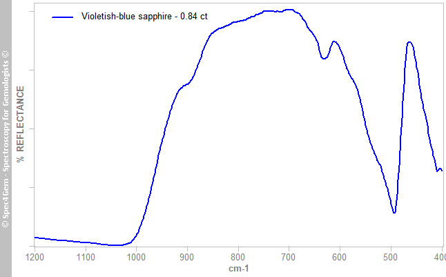
Figure 3. The IR reflectance spectrum acquired from the table, is closely related to the spectrum of the 0001 face of a corundum crystal.
UV-VIS-NIR spectroscopy:
The UV-VIS spectrum was acquired with a light path from culet to table, thus perpendicular to the table. With this orientation, the spectrum is the ordinary ray (E_|_c) spectrum.
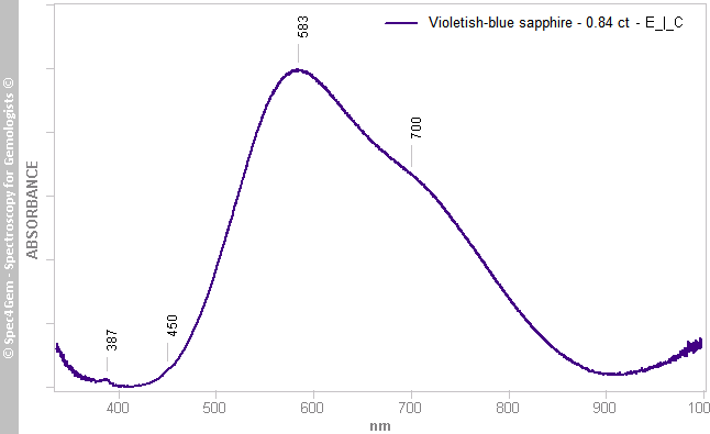
Figure 4. Ordinary ray (E_|_c) spectrum demonstrating the strong Fe2+-Ti4+ IVCT (583 and 700 nm) and weak features of Fe3+ (387 and 450 nm).
The ordinary ray (E_|_c) spectrum (figure 1) shows strong and broad absorption bands at 583 nm and 700 nm giving the blue color to the stone. Two additional features are present but rather weak at 387 nm and 450 nm.
The 583 nm and 700 nm absorption bands – in fact, the gaussian are centered at 567 nm and 684 nm – are attributed to the inter-valence charge transfer (IVCT) between the Fe2+-Ti4+ acceptor-donor pairs [2]. The narrow band at 387 nm is attributed to single Fe3+ ion although the narrow band at 450 nm is attributed to Fe3+-Fe3+ pairs exchange interactions but also to the spin-forbidden transitions of the single Fe3+ ions.
The Fe3+ absorption (386 nm and 450 nm) are rather weak, likely indicating a very low Fe3+ concentration. Such low Fe3+ concentration is quite uncommon in natural sapphire but it should not be forgotten this could also occur in mother nature. However, synthetic sapphires are generally containing just enough Fe2+ iron to cause the IVCT with Ti4+ in order to produce the strong blue color.
Photoluminescence spectroscopy:
The photoluminescence spectra are acquired from the table of the stone with the excitation ray parallel to the table entering the stone through the girdle.
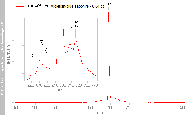 Figure 5. 405 nm excitation photoluminescence spectrum of the sapphire showing Cr3+ luminescence.
The luminescence of the stone with a 405 nm source shown in figure 5 is resumed to few features in the 700 nm region. The strong and main peak at 694 nm is consistent with Cr3+ R1-R2 emission lines in corundum. Few much weaker side peaks are also observed (view inset), 660, 671, 676, (685), 708 and 715 nm. The 676 nm peak (doublet) could be ascribed to Cr3+ in the chrysoberyl structure of corundum, as well as the 685 nm peak (doublet) could be ascribed to Cr3+ in boehmite structure of corundum ...
No orange luminescence is observed as it seldom is in sapphire originating from Sri-Lanka and Madagascar.
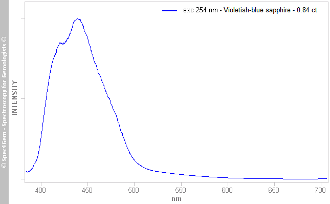 Figure 6. 254 nm excitation photoluminescence spectrum of the sapphire showing Ti4+ luminescence.
The luminescence of the stone with a 254 nm source was very weak, the excitation power of the source being low, a stronger source would have likely produced a stronger luminescence. Anyhow, the luminescence was observed with the spectroscope but it was invisible to the eye. In figure 6, the emission peak shape lets imagine two emission peaks, but this is likely the result of the noisy and low signal of the spectrum. Therefore, we consider a single emission peak centered around 430 nm with a width of about 80 nm. This is consistent with the data of Wong, W.C., McClure, D.C. et al. (1995a & b) [4], Richard W. Hughes & John L. Emmett [3], this emission peak can be attributed to Ti4+. It is important to note that this luminescence is usually observed in synthetic corundum produced at high temperature or heat-treated natural sapphires. Besides that, it not sure as of now that such luminescence does not appear in low temperature sapphire.
Discussion:
All results, classical gemology and spectroscopy, undoubtedly confirm the material is corundum (sapphire). The strong violetish-blue color is caused by the Fe2+-Ti4+ IVCT associated with a very low Fe3+ content. The material is also inert to a N52 magnet, belongs to the low RI range of corundum, confirming the low iron content. From that, it is not possible to definitely conclude this sapphire is of synthetic origin, but this is likely the case.
[1] Karl Schmetzer and Adolf Peretti. Some diagnostic features of Russian hydrothermal synthetic rubies and sapphires. Gems & Gemmology, 1999, Spring, pp. 17–28.
[2] Townsend M.G. (1968) Visible charge transfer band in blue sapphire. Solid State Communications, Vol. 6, pp. 81–83.
[3] Richard W. Hughes & John L. Emmett. Heat Seeker: UV Fluorescence as a Gemological Tool. http://www.ruby-sapphire.com
[4] Wong, W.C., McClure, D.C. et al. (1995a & b) Charge-exchange processes in titanium-doped sapphire crystals. Physical Review B, Vol. 61, No. 9.