
Green kyanites are quite rare especially the yellowish-green ones, the 10.15 ct sample presented here is reportedly from Mirerani (Merelani) in Tanzania. It should be noted that such a 'big' kyanite is not so common in such quality.
The material is not clean as it can be observed on figure 1. The table is rather rough, it is likely a crystal face that has been barely polished. Reading refractive indexes was rather difficult. No pleochroism was observed.
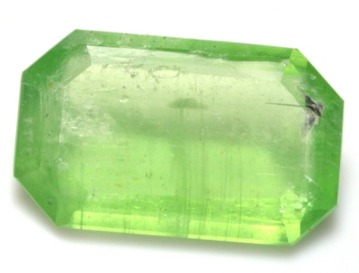 Figure 1. 10.15 ct yellowish-green kyanite reportedly from Mererani,
Tanzania
| Shape | 'emerald cut' |
| Size |
17.8 x 10.9 x 5.1 mm |
| Color | yellowish-green |
| Lustre | sub-vitreous |
| Weight | 10.15 ct |
| SG | 3.69 |
| RI | ~ 1.715 - ~ 1.732 |
| DR | ~ 0.017 |
| Pleochroism | unobservable or inexistant |
| Polariscope / Conoscope | light/dark 4 times / 360° |
| SWUV | inert |
| LWUV | inert |
| Magnetic susceptibility | weak with N52 |
Table 1. Observational and measured properties
Infrared reflectance spectroscopy:
The IR reflectance spectrum (figure 2), acquired from the table, exhibits a characteristic spectrum of kyanite specie.
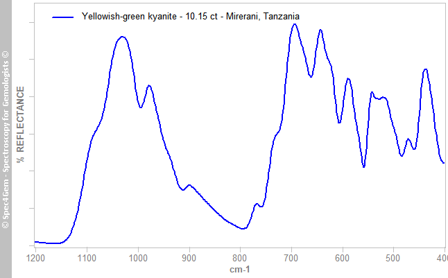 Figure 2. IR reflectance spectrum of the 10.15 ct yellowish-green kyanite from Mirerani, Tanzania, acquired from the table, exhibits the characteristic spectrum of kyanite specie.
 UV-VIS-NIR spectroscopy:
The UV-Vis-NIR absorption spectrum (figure 3) was acquired with the light path perpendicular to the table with the light entering the pavilion.
The unpolarized UV-Vis-NIR absorption spectrum exhibits features at 380, 433 and 446 nm related to Fe3+ as well as a broad band centered at 617 nm (Faye and Nickel, 1969)[2], (Parkin et al., 1977)[1]. Weak features unambiguously attributed to Cr3+ are also present at 691 and 709 nm. The Fe2+↔Fe3+ IVCT (Faye and Nickel, 1969)[2] is excluded for explaining the 617 nm band because the Fe2+ CF transitions absorption band in the 780-850 nm region (Faye and Nickel, 1969)[2] is not observed in this spectrum.
The 617 nm band has a comparatively low absorbance compared to the Fe3+ doublet at 433 and 446 nm, green and yellow are weakly absorbed / strongly transmitted, giving thus the yellowish-green color to the gemstone material.
A shoulder is present at 462 nm on the right hand side of the Fe3+ doublet. This shoulder is almost present in all kyanite spectra showing the Fe3+ doublet.
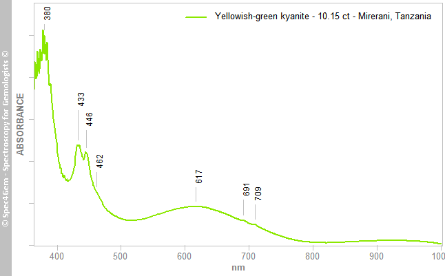 Figure 3. Unpolarized UV-Vis-NIR absorption spectrum of the 10.15 ct yellowish-green kyanite from Mirerani, Tanzania exhibits features at 380, 433 and 446 nm related to Fe3+ as well as a broad band centered at 617 nm. Weak features at 691 and 709 nm are attributed to Cr3+.
Two more spectra were acquired with polarizing filters that have a limited transmission bandwidth (420 - 800 nm). The collected spectra (figure 4) almost match the E||γ (green spectrum) and E||β (orange spectrum) directions. The E||β is slightly shifted for clarity. E||α spectrum was not acquired because of the long section of the stone that does not provide a light path easily workable even if it would have been possible anyway.
The Fe3+ doublet at 433 and 446 nm is invariable except its absorbance which is greater in E||γ direction. The evident Cr3+ features at 691 and 710 nm also depends on polarization, they are stronger in E||γ direction than in the E||β one. The 615 nm band in the E||γ polarization slightly shifts to 606 nm with a reduced bandwidth in the E||β polarization. A weak shoulder caused by another band seems to be present on the left hand side of the 615 nm band.
The weak difference between the E||γ (green spectrum) and E||β (orange spectrum) spectra explains that pleochroism is almost inexistent and was not observed.
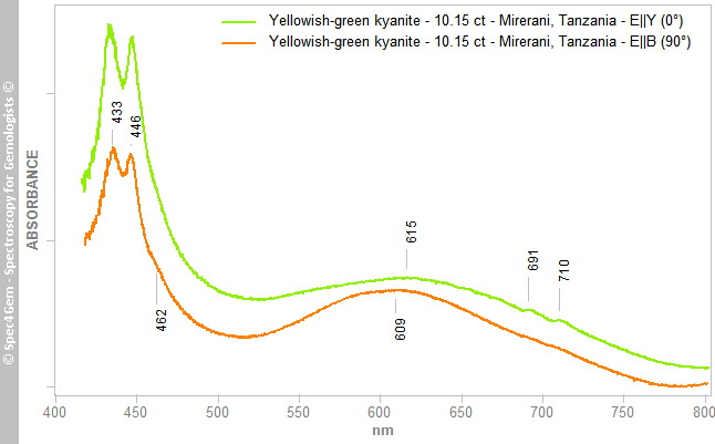 Figure 4. Polarized UV-Vis absorption spectra of the 10.15 ct yellowish-green kyanite from Mirerani, Tanzania for the E||γ (green) and E||β (orange) directions. The Fe3+ doublet at 434 and 446 nm is invariable except its absorbance increases in E||γ direction. The evident Cr3+ features at 691 and 710 nm also depend on the polarization, they are stronger in E||γ direction than in the E||β one. The 615 nm band in the E||γ spectrum shifts to 606 nm with a reduced bandwidth in the E||β spectrum.
Photoluminescence spectroscopy:
Although the gemstone is inert under the SW and LW lamps, a 405 nm laser pointer induces red luminescence. The photoluminescence spectrum (figure 5) excited with a 405 nm laser was acquired without any specific orientation.
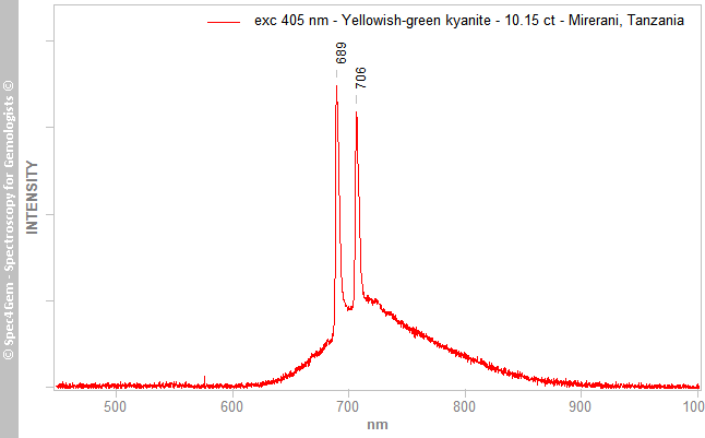 Figure 5. Photoluminescence spectrum of the 10.15 ct yellowish-green kyanite from Mirerani, Tanzania showing the Cr3+ R2 & R1 lines respectively at 689 and 707 nm.
This green kyanite shows a weak red luminescence attributed to the R2 & R1 lines of Cr3+ respectively at 689 and 707 nm (Gaft et al.)[5], these R lines are associated to the Cr3+ in site A. The 760 nm emission center (Cr3+ in site B) is also present although it does not clearly appear in the spectrum of figure 5. Using 635 and 678 nm lasers, the 760 nm emission center is just visible, luminescence is rather weak.
Conclusion:
The green color of this unusual 10.15 ct yellowish-green kyanite from Mirerani in Tanzania is due to Fe3+ ions. Even if Cr3+ is present as shown by the photoluminescence spectroscopy it likely has few or no impact on the color.
[1] Mineralogical Applications of Crystal Field Theory, R. G. Burns, 2005, ISBN: 0521017858 | 978-0521017855
[2] On the origin of colour and pleohroism of kyanite, G. H. Faye, E. H. Nickel, The Canadian Mineralogist, Vol 30, No 35, 1969
[3] Blue colour-changing kyanite from East Africa, G. Bosshart et al., Journal of Gemmology, Vol. 18, No. 3, pp. 205–212
[4] Kyanite Visible Spectra - List of Visible Data Files on the Caltech Mineral Spectroscopy Server
[5] Modern Luminescence Spectroscopy of Minerals and Materials, 2nd Edition, M. Gaft, R. Reisfeld, G. Panczer, Springer Editor, ISBN: 9783319247632