
Pink zoisite var. thulite from Tanzania reaching almost the carat
- Details
- Created: Sunday, 15 March 2020 10:58
Zoisite is very famous in jewellery because of its violet-blue variety named tanzanite after its country of discovery, Tanzania. Zoisite is the orthorhombic polymorph of clinozoisite, a member of epidote group. It occurs in many colors such as violet, blue, green, yellow, orange, red, pink, brown, gray and even almost colorless but among these colors some are rarer than others. Pink zoisite is quite common and is found in more than 150 locations over the world. However, Norway in the northern countries, is the main source for producing thulite cabochons and carvings. Thulite is the variety name of pink-red zoisite that has been in used for a long time to describe the massive stones where it is often mottled with calcite tiny crystals. Another definition of thulite is the pink zoisite that owes its color to Mn3+. Transparent crystal of thulite have been reported but published in the mindat's zoisite page[1], that is correct since thulite is a zoisite. The pink zoisites are sometime sold as pink tanzanite but this is inaccurate since tanzanite is only the violet-blue variety.
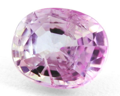 Figure 1. The 0.97 ct pink zoisite is badly cut with an excentric culet
and irregular shape. It hosts several inclusions planes and veils.
The trichroism makes the photography a bit tricky, with the naked-
eye the stone is pink but the camera is sensitive to the three colors
and their related direction.
The raised question is to know whether such a gemmy transparent pink zoisite can qualified for the thulite variety name or not. From former definitions, the gemstone is not thulite since it is not in the massive form but it is if colored by Mn3+. Similarly, the green beryl colored by Cr3+/V3+ is an emerald although a green beryl mainly colored by Fe2+ is nothing else than just a green beryl.
The pink zoisite studied here is reportedly from Tanzania and very likely from the Merelani area where the tanzanite and other zoisites are mined. Facetted 'fancy-colored' zoisites are sometime available from Pakistan but the palette of color is limited to few colors compared to that of Tanzania. A purplish pink crystal from Ariege in France was published in mindat[2].
| Shape | oval, irregular and culet is off-center |
| Size | 6.5 x 5.5 x 3.6 mm |
| Color | pink |
| Lustre | vitreous |
| Weight | 0.97 |
| SG | 3.13 |
| RI | 1.691 - 1.701 |
| DR | 0.010 B+ |
| Pleochroism | very strong: light-purple / red-violet (magenta) / light-green-yellow (see figure 2) |
| Polariscope / Conoscope | anisotropic: light/dark 4 times over 360° |
| SWUV | inert |
| LWUV | inert |
| Magnetic susceptibility N52 | diamagnetic |
| Chelsea filter | inert (same color as seen without filter) |
Table 1. Observational and measured properties
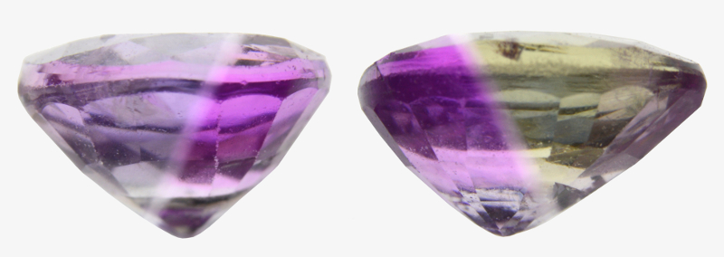 Figure 2. The very strong trichroism shows three colors: light-purple, red-violet (magenta), light-green-yellow.
Infrared reflectance spectroscopy:
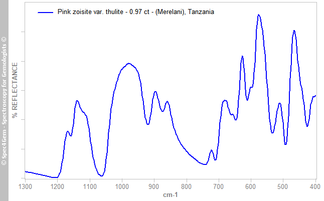
The infrared reflectance spectrum (figure 3) was collected from the stone's table and characterized the stone as zoisite. No investigation was conducted to look over the dimorphism of zoisite for determining if the stone is zoisite (orthorhombic) or clinozoisite (monoclinic).
UV-VIS-NIR spectroscopy:
Zoisite is trichroic and the studied stone does not depart from the general rule with its strong trichroism showing three colors: purple, light-pink and light-green-yellow. The UV-Vis-NIR spectra (figure 4) were collected by carrefully orienting the stone and using polarizing filters to separate the three polarization directions of the light through the crystal.
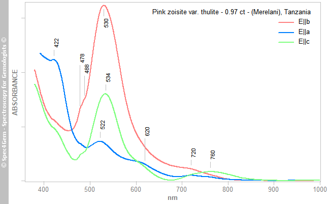 Figure 4. The Visible-NIR spectra collected from the three polarization directions show a great spectral anisotropy responsible for the strong dichroism. The bands are all ascribed to Mn3+ except the 620 nm one, which is related to V3+.
Even if all three spectra share common features at 422, 478/488, 522-534, 620, 720 and 760 nm, they significantly differ by the absorption bands intensity, explaining the strong trichroism. The 422, 522-534 and 720-760 nm bands are ascribed to the spin-allowed transitions of Mn3+ in the distorted octahedral M3 site, shoulder bands at 478 and 488 nm are ascribed to the spin-forbidden transitions of Mn3+. The 620 nm band is connected to V3+. The absorption edge towards higher energies below 400 nm is usually observed in V3+/Cr3+ zoisites.
In epidote structure, the Mn3+ ion (3d4) occupies the M3 site in octahedral coordination, a MO (Metal-Oxygen) site (Burns, 2005)[3]. The energy levels of the spin-allowed bands of the collected spectra are drawn in the energy level diagram of figure 5. The distorted octahedral site leads eg and t2g groups to split into additional energy levels with the possibility of spin-allowed transitions. This explains the three bands instead of the only predicted one (one spin-allowed transition) in an ideal octahedral configuration where Mn3+ is unstable. Each energy level correspond to the 3d orbitals (dxy, dxz, dyz, dz2, dx2-y2) and they are derived from the bands energies of the three polarized spectra by averaging them, they are relative to the eg ground state. The stabilization energy of the t2g orbitals split is δ1 that is calculated as follows: δ1 = 23,700 - 18,900 = 4,800 cm-1 and the stabilization energy of the eg orbitals split (caused by the Jahn-Teller effect) is δ3 that equals to 13,510 cm-1. The centre of gravity rules of the eg and t2g baricentres allow to calculate the crystal field splitting parameter Δo = 10Dq as follows: 23,700 - 2δ1/3 - δ3/2 thus Δo = 10Dq = 13,745 cm-1. It is consitent with the This Δo = 13780 cm-1 of the MK25 thulite sample (Langer et al., 2002)[4].
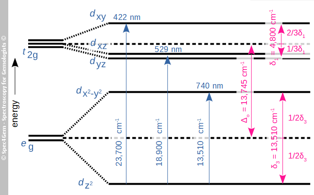 Figure 5. The energy levels diagram derived from the collected spectra with the three bands at 23,700, 18,900 and 13,510 cm-1 yields a crystal field splitting parameter of Δo = 10Dq = 13,745 cm-1.
Photoluminescence spectroscopy:
The photoluminescence spectrum (figure 6) was acquired with a 405 nm laser. Despite the power of the laser, the luminescence is rather weak. There is a weak emission at 469 nm related to REE3+ (Gaft et al., 2015)[5], the 693 and 712 nm weak emissions are related to V2+ (Gaft et al., 2015)[5]. The prominent 553 nm (18,080 cm-1) emission is possibly connected to Mn3+ but the lack of litterature on the subject and the unexplained / unassigned corresponding energy level do not allow a firm conclusion. Additionally the 447 nm laser induces the same luminescence emissions (469, 553, 693 and 712 nm) as for the 405 nm laser but comparatively they are so weak. The 532 nm laser does not induce any luminescence as well as the SW- and LW-UV sources. From that, the 553 nm emission requires excitation energies greater than 450 nm (22,300 cm-1) and lesser than 370 nm (27,000 cm-1). Mn2+ may also be present in comparatively low concentration compared to Mn3+, the emission wavelength and its broadness are compatible with Mn2+ as well.
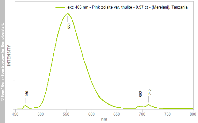 Figure 6. The 405 nm excitation photoluminescence spectrum shows a REE3+ emission at 469 nm, a possible Mn3+ emission at 553 nm and the V3+ emissions at 693 and 712 nm.
Conclusion:
This 0.97ct pink zoisite is undoubtly colored by Mn3+ as demonstrated by Visible spectroscopy. According to the thulite variety name definition, this gemstone can be qualified as 'zoisite var. thulite'. Is that naming so important ? In the trade, the thulite variety name has been in used for dacades and pertains to the massive form although the transparent pink gemstone is simply named pink zoisite.
[1] Mindat - Thulite crystals, the pink variety of zoisite, from the Terningmoen Military Camp, Elverum, Hedmark, Norway
[2] Mindat - Zoisite crystal (Visual id. possible clinozoisite), Petches, Ax-les-Thermes, Ariège, France
[3] Mineralogical Applications of Crystal Field Theory, R. G. Burns, 2005, ISBN: 978-521017855
[4] The crystal chemistry of Mn3+ in the clino- and orthozoisite structure types - A structural and spectroscopic study of some natural piemontites and thulites and their synthetic equivalents, K. Langer, E. Tillmanns, M. Kersten, H. Almen and R.K. Arni, Z. Kristallogr., 2002, 217 pp. 563-580
[5] Modern Luminescence Spectroscopy of Minerals and Materials, M. Gaft, R. Reisfeld, G. Panczer, 2015, Springer Editor, 2nd Edition, ISBN: 9783319247632